Imagine manufacturing a kidney, liver tissue, heart muscle or an arterial graft. Well that’s what researchers at University of Pennsylvania are doing using a 3D printer they built from an open source kit.
Bioprinting is a new science that allows tissue to be constructed using fabrication techniques. The challenge until now has not been in creating tissue but rather in developing 3D structures that include the vasculature needed to oxygenate and nourish the tissue.
The research team at University of Pennsylvania built a 3D vascular framework in the form of a lattice first and then added a gel consisting of human cells later. This was done in similar fashion to the way wax would be used in casting molds. The lattice, composed of three different sugars and a corn-based polymerase dissolved when water was flushed through the cellular tissue after it was applied. What was left was tissue with all the vasculature in place to ensure viability.

The use of sugar provided the researchers with a best fit with other organic material. Sugar proved to be compatible with 3D printing technology as well. The formula consisted of sucrose, glucose and dextran. The printer used was the RepRap.

Although most 3D printers create models using a layering approach, the University of Pennsylvania team, led by Jordan S. Miller, Christopher S. Chen, working with Sangeeta N. Bhatia and Kelly R. Stevens of MIT, built a 3D lattice of cylindrical filaments first. Then they applied to the lattice a gel containing living human cells including blood vessels. Injected water dissolved the sugar and then fluids were introduced to feed the cells nutrients and oxygen. The human blood vessel cells spontaneously generated new capillaries in the same way blood vessels grow in the human body.
Still to be done: learning to connect artifically created vascular networks with living networks in the body.
But the implications of this technology are enormous. Instead of using transplants and having to deal with potential rejection or a lifetime of anti-rejection medications, we will be able to have our own cells extracted and cultured to grow replacement parts for diseased organs and damaged tissue.
The researchers recently published their results in Nature Materials.
Related Posts
 Canada is Becoming a Nuclear Fusion Hotbed
 The Nuclear Arms Race Never Ended
 If You Are Using Semaglutide Drugs to Lose Weight Are You Hooked on Them for Life?
 Weaponized Mosquitoes Are Being Deployed to Reduce Vector-Borne Diseases
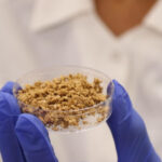 Transforming Coffee’s Future With Bioreactors And Cell Power
 Can Planets Exist Without a Star?
 Conversations About AI: Will Human Software Coders Soon Be Obsolete?
 Have We Learned Anything From The COVID-19 Pandemic?


Does your site have a contact page? I’m having a tough time locating it but, I’d like to send you an email. I’ve got some ideas for your blog you might be interested in hearing. Either way, great blog and I look forward to seeing it grow over time.