
Until COVID-19, I had no heart issues. On the other hand, my daughter, who is 40 now, was born with multiple heart defects. Because of her struggles, I have become very familiar with the vital organ in our bodies that keeps us going for an entire lifetime.
I am 76 now, and my post-COVID heart condition is under control. My daughter, who has had multiple heart operations, is doing well and has a four-year-old daughter. All of this good news hasn’t stopped me from continuing to research and write about the heart.
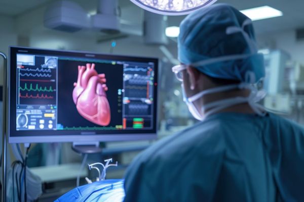
In this posting, I look at two encouraging stories. The first is about a University of Texas at Arlington (UTA) invention of a 3D-printed, bioengineered heart patch that regenerates damaged heart muscles. The second is a Columbia University (CU) invented artificial intelligence (AI) tool that can more accurately detect structural heart issues by interpreting electrocardiograms (ECGs).
New Smart Bioengineered Heart Patch
A polymer patch containing exosomes has been developed at UTA to address inflammation and stimulate heart tissue regeneration. The patch is a flexible material that adapts to the contractions of the heart muscle and conducts electrical impulses. It is 3D printed and, unlike scar tissue that forms after a heart attack, mimics natural cardiac tissue at both micro and macro levels.
Yi Hong, in the UTA Department of Bioengineering, has been working on a bioengineered 3D-printed solution to support heart structure and promote regeneration after a heart attack. He has collaborated with Ke Cheng at CU in the development of their current invention.
The keys to the patch are two-fold. It is a flexible polymer, and it is embedded with exosomes. Exosomes are tiny structures produced in cells and then expelled into the surrounding environment, where they can be found in fluids like blood, saliva, cerebrospinal fluid and urine. They act as messengers between cells and deliver lipids, DNA, mRNA and microRNA to surrounding cells, helping to repair and regenerate tissue. Exosomes also remove unnecessary molecules, acting like a waste disposal system. That’s what makes them highly suitable for the treatment of damaged heart muscles. The patch releases exosomes for cardiac tissue regeneration in treating heart attacks and represents a promising new therapy.
AI App Detects Heart Structural Defects From ECGs
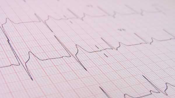
I regularly do a 30-second ECG using my KardiaMobile app. It can detect up to six of the most common arrhythmias (a fancy word for abnormal rhythm patterns). I subscribed to KardiaMobile after I was diagnosed with atrial fibrillation (AFib) after contracting COVID-19.
ECGs are the first test that doctors and cardiologists use when checking the heart. They are great at detecting electrical problems in the heart, but are not commonly used to determine if there are structural defects in the organ. For the latter, echocardiograms (ultrasound pictures of the heart muscle and blood vessels) are the first test of choice.
A research team at CU wanted to see if they could use the ECG as a more effective and cheaper screening method for detecting structural heart defects. They created an AI tool called EchoNext and published their research on July 16, 2025, in the journal Nature. The AI can look at a standard ECG and single out patients who show subtle variations in the readings to suggest a need for an ECG.
Effectively, EchoNext makes the much cheaper test a far more effective screening tool for finding structural heart defects that cardiologists would not see just from reading an ECG. EchoNext has been trained using more than 1.2 million ECGs done on 230,000 patients. In validation testing, it has been proven to accurately identify structural heart defects, including cardiomyopathy, valve disease, pulmonary hypertension, and severe thickening of the heart muscle. When challenged in head-to-head comparisons with 13 cardiologists looking at 3,200 ECGs, EchoNext proved it could find structural heart problems that the doctors missed. In a further test with 85,000 patients, EchoNext identified over 7,500 individuals at high risk for undiagnosed structural defects. Of those, 55% had subsequent echocardiograms, with 75% being diagnosed with structural heart disease. This was twice the rate of identification when compared to patients having a first ECG without EchoNext.
States Pierre Elias, Assistant Professor of Medicine and Biomedical Informatics at CU’s Vagelos College of Physicians and Surgeons, and Medical Director for AI at NewYork-Presbyterian, “You can’t treat the patient you don’t know about. Using our technology, we may be able to turn the estimated 400 million ECGs that will be performed worldwide this year into 400 million chances to screen for structural heart disease and potentially deliver life-saving treatment at the most opportune time.”