
Ever since my daughter was born in 1984 and was diagnosed with a congenital heart disease (CHD), I have been researching and writing on the subject.
My daughter was four before she underwent open-heart surgery to repair her CHD, Tetralogy of Fallot (ToF) with Absent Pulmonary Valve. The surgeon patched the large hole between her two ventricles, the lower pumping chambers of her heart. Then a homograft pulmonary valve (a valve harvested from a human donor) was inserted between the right ventricle and pulmonary artery leading to her lungs.
Numerous hospitalizations followed. At age 8, she had a second open-heart operation to replace the homograft valve. A third operation followed at age 24. Today, she is 42 with a 5-year-old daughter. She has had a good outcome, with the likelihood of her needing another valve replacement years down the road.
Autus: A New Heart Valve for Children with CHD
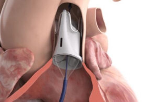
A clinical trial involving 62 children in 12 different U.S. locations has led to the U.S. FDA approving Autus, a new size-adjustable heart valve for patients with Tetralogy of Fallot, the CHD my daughter had at birth. She had an added complication. The pulmonary valve was missing, causing lung complications beginning with her first breath.
Autus is adjustable. If implanted in a toddler with its initial manufactured diameter of 12.7 millimetres (0.5 inches), it can serve the child well for a time. Then, as the child grows, Autus can be expanded in 2 millimetre increments to eventually reach 22 millimetres (0.866 inches) by the time the child becomes an adult.
In past heart valve surgical procedures on children, often an adult-sized valve is the only size available. This means the valve has to be adjusted to fit the child, leading to multiple open-heart and valve replacement surgeries. With Autus, however, it can be optimized to the child’s body size in one operation and then be adjusted as the child grows to adulthood. Each 2 millimetre adjustment involves the far less invasive procedure that my daughter experienced at age 24.
What Makes Autus Different?
Besides the ability to adjust the diameter of the valve, Autus uses a polymeric material for its valve leaflets, the first of its kind. The polymeric material is a synthetic fluoropolymer membrane that performs like normal heart valve tissue to regulate blood flow from the heart to the lungs. After one year, the clinical trial follow-ups showed no valve leaflet wear.
Autus can replace prosthetic mechanical, homograft, porcine or bovine valves. This means a better quality of life for children with CHD, and fewer traumatic surgical interventions and long periods of recovery.
The Autus framework is constructed from stainless steel. It includes a polymeric sleeve to allow Autus to be secured to the surrounding pulmonary outflow tract tissue leading from the right ventricle into the artery and the lungs. It is the first polymeric leaflet valve to be approved by the FDA.
During FDA testing, the Autus was subjected to the equivalent of 15 years of heartbeats. Its inventor, Dr. Sophie-Charlotte Hofferberth, told Medical Design & Outsourcing, “Because of that size adjustability, it could certainly last for longer than that,” referring to the 15-year testing and 600 million cycles of frame fatigue Autus endured from FDA reviews.
Dr. Hofferberth and her partners, Dr. Elazer Edelman and Pedro del Nido, invented the valve and founded their company, Autus Valve Technologies, in 2019. Autus was acquired by Edwards Lifesciences this year.
Facts About Congenital Heart Disease
The word “congenital” means present at or from birth. Congenital diseases can be genetic or occur when something happens during pregnancy or at the time of birth.
CHDs vary from moderate to severe and occur in 1% to 1.2% of live births. Approximately 1.35 million children are born with CHD annually. Some CHDs can go undetected into adulthood. Others, however, can be immediately life-threatening.
A list of the most common CHDs follows:
Atrial Septal Defect (ASD)
This is a hole in the wall (septum) that separates the heart’s atria, the two upper pumping chambers. This compromises normal blood circulation.
Ventricular Septal Defect (VSD)
This is a hole in the septum separating the ventricles, the heart’s two lower pumping chambers. This also compromises normal blood circulation.
Patent Ductus Arteriosus (PDA)
Its cause is the failure of a necessary fetal blood vessel called the ductus arteriosus to close after birth. This leaves an abnormal connection between the aorta and pulmonary arteries and impacts blood circulation. A persistent PDA can mask symptoms of other CHDs.
Patent Foramen Ovale (PFO)
This CHD is a flap that covers an opening between the two atria. Normally, it fuses shut. When it doesn’t, its presence can lead to ischemic strokes or a transient ischemic attack (TIA).
Pulmonary Stenosis (PS)
This involves narrowing of the pulmonary artery and the obstruction of blood flowing from the right ventricle to the lungs. It causes the right ventricle to work harder and thicken over time.
Coarctation of the Aorta (CoA)
This describes a narrowing of the aorta, the main artery that delivers blood and oxygen to the body. It causes the left ventricle to thicken and lead to heart failure.
Bicuspid Aortic Valve (BAV)
The aortic valve has only two valve leaflets rather than three. It can work for years but can begin to leak. Leaky heart valves are never a good thing.
Tetralogy of Fallot (TOF)
This rare CHD combines four heart defects: a VSD, PS, an abnormally positioned aorta, and right ventricle wall thickening (hypertrophy). When the pulmonary valve is absent, it can severely damage the lungs as they begin to inflate after birth. It is one of several CHDs referred to as blue baby syndromes (cyanosis).
Transposition of the Great Arteries (TGA)
This CHD involves a structural defect involving the aorta and pulmonary arteries, which are connected to the wrong ventricles. This creates two separate blood circulation paths. Oxygen-rich blood circulates between the heart and lungs. Oxygen-poor blood only circulates between the heart and body, leading to cyanosis.
Hypoplastic Left Heart Syndrome (HLHS)
This CHD involves the underdevelopment of the left side of the heart, which imperils blood flow from the lungs and heart to the body, causing cyanosis.
Atrioventricular Septal Defect (AVSD)
This condition involves the abnormal development of the walls that separate the heart’s four chambers, with abnormal development of two heart valves, the mitral and tricuspid. It causes cyanosis.
Hypertrophic Cardiomyopathy (HCM)
This involves an abnormal thickening and stiffening of the heart muscle, which can show up much later in life, leading to sudden cardiac death. My father-in-law and his sister both had HCM. He had a pacemaker implanted and died at age 97. She was 102 when she passed away.