
I tend to seek out information about innovations related to the cardiovascular system. I attribute this to having a daughter born with congenital heart disease and the more recent impact on my heart from my first encounter with COVID-19.
Cardiologists have a suite of tools designed to monitor heart function. Ultrasounds, also known as echocardiograms, are used to get a look at the physical heart using sound waves. An ultrasound can tell a cardiologist a lot. Are the four chambers normal in appearance? Are the valves functioning properly? Are there leaks? Are they seeing normal blood flow? Is the heart muscle contracting as it should?
To detect irregularities in the heart’s conduction system, electrocardiography is used. This involves getting tracings of the electrical signature of all four chambers and the major blood vessels. When done in a medical office, leads are applied to various areas of the body, upper chest, abdomen and legs. A more portable approach is to use a Holter Monitor which can be worn externally for a number of days to provide a complete record of every beat and missed beat. Then there are portable devices like the Apple Watch and the one I use, Kardiamobile, a pad that combines with a smartphone app to give me a 30-second electrocardiogram which I can then send to my cardiologist. If I have an odd feeling in my chest, I use the pad which is split in two, with two fingers pressed on each side. Since being diagnosed with atrial fibrillation (aF) I tend to use it at least once a day or anytime I feel light-headed or have odd palpitations in my chest.
In addition to the ultrasound imaging previously described, heart function can also be assessed by an MRI, CT scan, and X-rays. More invasive are cardiac catheterization procedures which involve inserting a catheter into a blood vessel and threading it into the heart where a contrast dye and X-ray provide a good two-dimensional real-time image of the heart as it works.
But what if you could put a patch on your skin and use it to monitor heart function? Up until now, wearable devices that used leads to contact the skin, or watches were the only ones capable of producing heart data useful to medical practitioners. These were exclusively focused on the heart’s conduction system. But with a new invention from the University of California San Diego, these may soon be joined by a companion wearable the size of a postage stamp that could become the way ultrasonic images of the heart are taken.
The patch provides accurate continuous monitoring of cardiac performance using ultrasound images. The illustrations below come from an article published in the January 25, 2023 edition of the journal Nature.
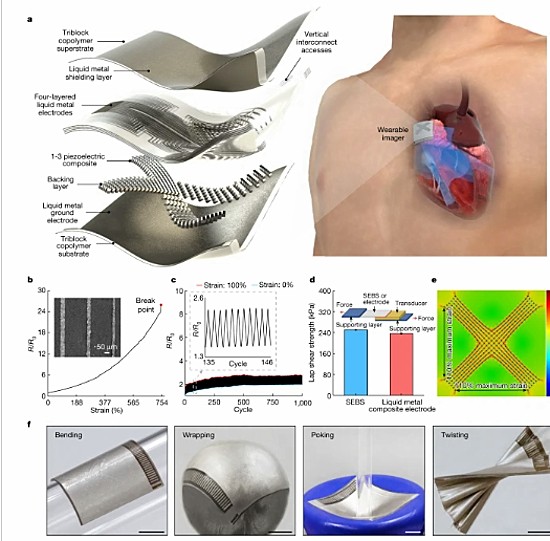
The wearable patch contains liquid metal composite electrodes and piezoelectric transducers. It can image deep into the body and provide a range of images including axial, elevational, and lateral views. Compared to regular ultrasounds the differences when looked at by third parties were considered negligible.
Unlike regular echos, the images this patch can gather can see the heart when a patient is in motion rather than lying on an examination table. Ultrasounds cannot image the image when it is under stress, only after a stress test when the patient is then examined. Conventional ultrasounds use gels which are critical to being able to obtain images. Gels are constantly reapplied throughout an imaging session. The patch uses liquid silicone which retains its liquid state for a full 24 hours so that images can continuously be gathered.
The developers have also created deep learning modelling software which when combined with the images provides measurements of stroke volume, cardiac output and ejection fractions making this technology not only versatile but also likely the next tool in a cardiologist’s arsenal.