March 14, 2020 – Today is Pi day because when the date is written as 3.14.2020, the first three numerals represent the value Pi the ratio of a circle’s circumference to its diameter. Take the Earth for example. The circumference at the equator is 40,075 kilometers (24,901 miles). The diameter is 12,742 kilometers (7,917.5 miles). Divide the circumference by the diameter and you get 3.14 followed by a string of numbers. My son-in-law proposed to my daughter on Pi Day because he and many other nerds celebrate this mathematical ratio reflected in the calendar. Guess what is his favourite dessert? No doubt you have figured out it is pie.
It is strange how our brains latch on to ideas that become lifetime fixations like Pi Day. And the more we study the brain, the more we realize just how marvelous an organ it is. Today when we want to study the brain using imagery we are largely limited to MRI, CT and PET scans. These technologies are complex and capital intensive.
But that may soon end if the work being done at Imperial College of London, and University College London moves beyond a current proof-of-concept to become a standard new imaging technique. The researchers at these two institutions have invented a small device that can be placed on an ambulance to provide brain scans in situ, giving neurosurgeons and emergency health teams literally “a heads up” on a traumatic injury before arriving at the hospital.
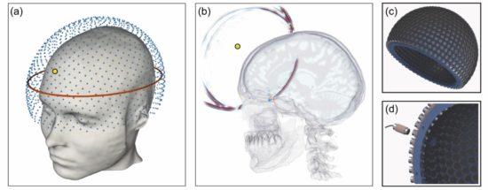
This technology uses seismic waves to create a detailed image of the brain. The device resembles a helmet and Bryan Williams, a professor and Director of the National Institute of Health Research at University College London describes the invention as “an extraordinary and novel development in brain imaging which has huge potential to provide accessible brain imaging in routine clinical practice to evaluate….head trauma, stroke and a variety of brain diseases…If this lives up to its promise it will be a major advance,” and is “a fabulous illustration of how the collaboration between engineers and clinicians, using methods from another sphere of science, can bring ground-breaking innovation into medical care.”
To what disciplines is Williams referring? Geologists and petroleum engineers use seismic data and a computational technique called Full Waveform Inversion (FWI) to map the inside of our planet. Seismic data picked up from strategically placed seismic sensors measure Earth movement. When plugged into FWI algorithms they can create three-dimensional images of the Earth’s crust, useful for oil and gas exploration, and for studying earthquakes. Now FWI is being applied to studying the brain producing high-resolution images.
The helmet is lined with an array of acoustic transducers that send sound waves through the skull and into the brain. The recording produced is analyzed using FWI to create a detailed image. With test subjects using the prototype, the researchers have been able to create detailed images of the brain that can be studied for signs of a stroke, the second most common cause of death and the most common condition associated with adult neurological disability.
The advantage of the FWI device over MRI, the latter the current gold standard for detailed imaging of the brain, is the former’s portability. The FWI helmet can work anywhere, whereas MRI requires a dedicated hospital space and a controlled environment. An MRI cannot be used on patients with metal implants whereas the FWI can.
States Parashkev Nachev, a professor at University College London, and co-author of the study describing the invention states, “This is a vivid illustration of the remarkable power of advanced computation in medicine. Combining algorithmic innovation with supercomputing could enable us to retrieve high-resolution images of the brain from safe, relatively simple, well-established physics: the transmission of soundwaves through human tissue…Neurology has been waiting for a new, universally applicable imaging modality for decades: full-waveform inversion could well be the answer.”
A paper published on March 6, 2020, in Nature’s npj Digital Medicine journal describes the device in greater detail.
